2012年11月24日星期六

Observation 4 --- Week 6 (Extra observation on 20th Nov)

Because of missing one observation in week 2, I appreciate that our instructor Mr Mcfarland allows me do an extra observation on 20th Nov. In this observation, i have seen  lots of organisms appears in past observation. of course, more new organisms still can be find, and this time i find some other diatoms which really has a interesting shape.

1.melosira
other than the rectangle shape fragellaria i found in last observation, melosira is another diatom that have a geomatric shape. This square shape substance has a golden color under microscope and keep stationary for the whole time.
  Fig 17.1
if we see closely , we can see substance moving within the square. the figure below provides closer look. 
 Fig 17.2


Referance:

    Canter-Lund H and Lund J.1995. Freshwater Algae. Bristol: Biopress Limited. Fig 250-252, 138 p.

2.gomphonema 
This is another diatom. it does not have a very rigid shape as melosira and fragellaria. it likes to attach to the plant, but I found this one stay alone by itself. it also have a golden color under microscope. 
Fig 18.1
if we see closely enough , we can also see substance moving within itself. the figure below provides closer look. 
Fig 18.2 

Referance:

    Canter-Lund H and Lund J.1995. Freshwater Algae. Bristol: Biopress Limited. Fig 230-231, 128 p.

 3. Euglena
 This organism appears as a black dot under microscope, very small and move slowly.i found it near the plant. 
 Fig 19.1
 Fig 19.2
 Referance:
    Patterson DJ.1992. Free-Living Freshwater Protozoa. Washingon DC: ASM Press. Fig 118-122, 61 p.

 4. Raphidiophrys
 this orgamism has a round shape but have lots of flagella point out from the inside of this substance. it doest not move and i found them around the dirt or near the plant. 
 Fig 20.1
 Here is a closer look of this organism. 
 Fig 20.2

   Referance:
    Patterson DJ.1992. Free-Living Freshwater Protozoa. Washingon DC: ASM Press. Fig 405-408, 173 p.
 5. Unkown substance
 This is just a substance having irragular shape, and during the observation, i can see smaller organism moving from the irragular shape substance to the round unknown organism. I ask Mr McFarland about it, he was not sure about it either. 
 Fig 21

2012年11月19日星期一

Observation 3 --- Week 5 (observation time: 16th Nov 2012)

In this weeks, i am still be lucky to find new organisms. however a lots of the organisms starts to die. i can see lots of reains of organisms on the glass. This time i am only focus on a region only which is near the soil at the bottom.

 1. The organisms i find in the past observations 

(Eucblanis,Vorticella and Blepharisma)

 I am not going to give further explaination of these three organism, please refer to my previous two observations.

Eucblanis

  Fig 12.1

The picture below the dead Eucblanis, we can see that it does have a very tough outer layer, and the tail. 

 Fig 12.2

 Vorticella

 Fig 12.3

Blepharisma

  Fig 12.4

The the follow few organism is what i find new in this observation

2.Chrysamoeba 

 Fig 13

this organism does not move a lot, and within itself we can see lots of vacules too. it has very unique shape as well.  

Referance:

    Patterson DJ.1992. Free-Living Freshwater Protozoa. Washingon DC: ASM Press. Fig 140, 75 p. 
3.Fragellaria 
 Fig 14.1
this is a rectangle substance has a goldish-yellow color under microcsope in this observation. it is almost everywhere at the bottom. the picture below brings a close look of this substance. 
 Fig 14.2

Referance:

    Rainis KG and Russell BJ.1996. Guide to Mirolife. New York, London, Hong Kong and Sydney: Franklin Watt Publisher. Fig 43, 113 p. 


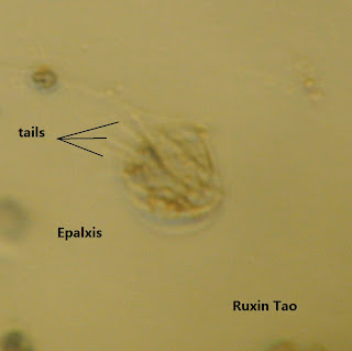
4.Epalxis
  Fig 15.1 
 this organism looks like a smaller version of jelley fish, it swims fast with its tails, it is not easy to take a picture under microscope. its shape are alsos very unique and can be easily identify from other organism.
 Fig 15.2
 Referance:
    Patterson DJ.1992. Free-Living Freshwater Protozoa. Washingon DC: ASM Press. Fig 311, 141 p.  

5.Lepidodermella 
 Fig 16.1
this is also a snail-like organism, moves freely in the water with two tails at the back. just like Blepharisma, it also has lots of vacules in itself to store food. pictures below is a close look of this organism when it is turning to another direction. 
 Fig 16.2
Fig 16.3


Referance:

    Rainis KG and Russell BJ.1996. Guide to Mirolife. New York, London, Hong Kong and Sydney: Franklin Watt Publisher. Fig 79, 177 p. 

2012年11月12日星期一

Observation 2 --- Week 4 (observation time: 9th Nov 2012)


In this observation, i find some of new organisms and also observe some from the first observation and compare the differences between these two observation. 


1. Chironomus 
this is what i see one week later, on 9th Nov i am lucky to find an live leaca of Chironomus
 Fig 3.3
 for the larva, it moves fast and it likes to flip its body from tail to head almost at the same position, the picture below was what i took at the same position
 Fig 3.4


Referance:
Ward HB, Whipple GH. 1918. Fresh-Water Biology. New York: Jonh Wiley & Sons, INC. Fig 1384, 915p.  

2.Bursaria 

 this is also an organism moves rapidly around, this is the picture of it near the plant

 
Fig 8.1
The picture below shows Bursaria doing some exchange with other organism 
 Fig 8.2
Then, we take a close look of this organism, you can see the three or more tails at the back of this organism
Fig 8.3
 Referance:

    Patterson DJ.1992. Free-Living Freshwater Protozoa. Washingon DC: ASM Press. Fig 318, 143 p.


3.Ameoba

this is an organism having very irregular shape, see the picture below

 Fig 9
I make a small movie to show how Ameoba exchange substance in or out of itself


 Referance:

    Patterson DJ.1992. Free-Living Freshwater Protozoa. Washingon DC: ASM Press. Fig 194-197, 198 p. 

4.Blepharisma

this is a snail-like  organism, it moves relatively slow. It likes to live near the soil. 
   
Fig 10.1
we can see the inside structure clearly for this organism,  lots of food vavuoles inside. 
 Fig 10.2


Referance:

    Patterson DJ.1992. Free-Living Freshwater Protozoa. Washingon DC: ASM Press. Fig 324-326, 146 p.   

5.Arcella

this is a single cell organism, it keeps stationary most of time when i observe it under microscope. however, when i take this picture, i find there this something attach on the Arcella, and both of them have identical shape. i have no idea what is it, but i think it maybe just something come out from Arcella or Arcella just doing the cell division.
 Fig 11


Referance:

    Patterson DJ.1992. Free-Living Freshwater Protozoa. Washingon DC: ASM Press. Fig 171-173, 91p. 

2012年11月5日星期一

Observation 1 --- Week 3 (observation time: 2nd Nov 2012)

I miss the first week's observation, therefore my first observation is about 12 days later from the start, on 2nd Nov, and there are huge changes within these 12 days.
Fig 3 
   
1.Chironomus 
From the picture we can see there is a insect at the up right coner, lets have a closer look 
 
Fig 3.1

   I lost this insect when i was doing my observation under microscope. however i found out the remains of the larva of this insect
Fig 3.2

 Referance:
Ward HB, Whipple GH. 1918. Fresh-Water Biology. New York: Jonh Wiley & Sons, INC. Fig 1384, 915p.

2.Anisonema

 During the observation, i found a interesting single cell organism with two tails at each side of the cell. It moves in a normal speed, therefore i am able to get clear pictures under microscope.

   
Fig 4.1

I also got another picture the two Anisonema hold together, i guess they may have some kind of comunication between. see Fig below 
Fig 4.2  

Referance:


    Patterson DJ.1992. Free-Living Freshwater Protozoa. Washingon DC: ASM Press. Fig 78, 54p. 


3.Vorticella

This is bell shape single cell organism attaching to the plant stem, it is Vorticella, they always comes with several individual together. see fig below
Fig 5.1

 Let's get a closer look of this organism. This organism almost keep stationary all time, the bell head may shake a little bit according to the water flows nearby.
 Fig 5.2

 Referance:

    Patterson DJ.1992. Free-Living Freshwater Protozoa. Washingon DC: ASM Press. Fig 232-236, 113 p.

4.Eucblanis

Eucblanis is the very unique organism i found in the observation. it moves in a fast speed with the help of its tail. 

 Fig 6

Referance:

Smith DG. 2001. Penna's Frashwater Invertebrates of The United States. New York: John Wiley & Sons, INC. 131p.  


5.Coleps

From the Larva of the Chironomus, i observed a gourp of single cell oganisms moving rapidly in side the remains of the Chironomus. Below is the short vedio i recorded

blow is the simple coleps i found from the microscope, this organism is moving very fast, but sometimes it will just keep stationary for a second or two. 
  
Fig 7

 Referance:

    Patterson DJ.1992. Free-Living Freshwater Protozoa. Washingon DC: ASM Press. Fig 375-377, 162 p. 


2012年10月29日星期一

Set ups --- Week 1

                                                                       Fig 1


           start of the project:
 
   we are trying to set up an environment for algea and other organism. The algea is used for provide food and oxygen to the organism. In addition , we use different water souce for this experiment, from different water souce we will have different organism with it. I use the following souce:

3. CARTER MILL PARK AT SPRING SOURCE


   3. Carter Mill Park at spring source   
 Fig 2
Carter Mill Park at spring source, Carter Mill Road, Knox Co. Tennessee Partial shade exposure N36 01.168 W83 42.832 940 ft 10/9/2011 


source from:  Botany 111 Fall 2012 Blog,web http://botany1112012.blogspot.com/

Here is the picture for the close look of the plants:

     Fig 3

Plant A . Amblestegium sp. Moss. Collection from: Natural spring. at Carters Mill Park, Carter Mill Road, Knox Co. TN. Partial shade exposure. N36 01.168 W83 42.832. 10/9/2011 

Plant B. Utricularia gibba L. Flowering plant.A carnivous plant. Original material from south shore of Spain Lake (N 35o55 12.35" W088o20' 47.00), Camp Bella Air Rd. East of Sparta Tn. in White Co. and grown in water tanks outside of greenhouse at Hesler Biology Building. The University of Tennessee. Knox Co. Knoxville TN. 

source: Botany 111 Fall 2012 Blog, web http://botany1112012.blogspot.com/